

Mandibula, яъне устухони ҷоғи поён, танҳо сохтори пассиве нест, ки дандонҳоро нигоҳ дорад. Он дар бисёр вазифаҳо, аз ҷумла хоидан, сухан гуфтан, нигоҳ доштани сохтори рӯй ва дастгирии дандонҳо ва implant-ҳо иштирок мекунад. Вақте mineral density ва microstructure-и ҷоғ заиф мешавад, дастгирии дандон, implant stability, orthodontic anchorage, periodontal health ва resistance ба jaw fractures метавонанд зарар бинанд.
Яке аз нишондиҳандаҳои муҳими bone health bone mineral density мебошад. Бо истилоҳи англисӣ bone mineral density ё кӯтоҳ BMD, ин basic measure-и вобаста ба mineral content-и bone мебошад. Аммо bone quality танҳо бо BMD шарҳ дода намешавад. Structure-и trabecular bone, trabecular thickness, number, spacing, connectivity ва organization низ mechanical strength-и bone-ро таъсир медиҳанд. Аз ин рӯ study на танҳо bone density, балки mandibular microarchitecture-ро ҳам арзёбӣ кардааст.
Starting point-и research ду mechanical stimulus-и гуногун аст. Якум systemic physical activity, мисли running. Дуюм local mechanical load, ки ҳангоми chewing мустақиман ба jaw bone мерасад. Previous studies нишон додаанд, ки exercise метавонад BMD-ро махсусан дар weight-bearing bones мисли femur зиёд кунад. Аммо mandibula аз ин viewpoint bone-и more complex аст. Developmental ва biomechanical properties-и он аз long bones фарқ мекунанд; он cyclic forces аз chewing muscles мегирад ва only movable bone of facial skeleton мебошад. Аз ин рӯ effect-и exercise on mandible clear нест.
Second problem-и study regional effect-и masticatory load мебошад. Hard food requires greater chewing force. Ин load дар mandibula махсусан around molars concentrate мешавад. Аммо whether anterior region responds similarly unclear аст. Therefore research mandibula-ро not as single unit, but separately as molar region and anterior region таҳлил мекунад.
Чаро ин problem important аст?
Барои dentistry, implantology ва maxillofacial surgery mandibular bone quality critical мебошад. Osseointegration of implant, stable tooth support, progression of periodontal disease ва response to orthodontic forces ба density ва microarchitecture of jaw bone вобастаанд. Агар mandibular bone weak бошад, implant stability decrease, bone loss accelerate ё treatment planning more risky шуда метавонад.
Аз perspective-и daily life, study ду behavior-и apparently simple-ро scientifically questions мекунад: moving ва chewing. Одамон generally медонанд, ки exercise барои leg, hip ё spine bones useful буда метавонад. Аммо whether jaw bone also affected by systemic exercise less discussed аст. Likewise, soft-food diet метавонад mechanical stimulus to chewing muscles and jaw bone-ро reduce кунад; аммо which mandibular regions show clear effect requires detailed study.
Study муҳим аст, зеро exercise ва masticatory load-ро separately evaluate мекунад. Aerobic exercise systemic stimulus мебошад; он whole skeleton-ро through blood flow, hormones, bone formation-resorption balance ва metabolic signals affect карда метавонад. Masticatory load local mechanical stimulus мебошад; direct-loaded area bone adaptation-ро trigger карда метавонад. Research кӯшиш мекунад нишон диҳад, ки these two mechanisms same ё different effects on mandible доранд.
Research кадом gap-ро address мекунад?
Existing literature effect of exercise on femur ва other weight-bearing bones-ро бештар омӯхтааст. Effect on mandible less clear аст. Mandibula intramembranous ossification арқылы develops ва within facial skeleton distinct biological/mechanical properties дорад. Thus exercise effect observed in femur cannot be assumed identical in mandible.
Masticatory-load literature ҳам similar limitation дорад. Animal studies show changes in chewing force can affect alveolar-bone microstructure. Аммо studies mostly focus on molar grinding region. Anterior tooth region has different loading due to cutting/gnawing. Therefore whether masticatory load produces same bone gain in anterior mandibular region is separate question.
Research examines both exercise intensity and diet-hardness-related masticatory load in same experimental framework. Moderate and high intensity exercise compared шуданд; effects of soft and normal-hard diets on femur, mandibular molar region ва mandibular anterior region separated шуданд.
Experimental animals ва groups
Study 24 3-week-old male Sprague-Dawley rats истифода кардааст. Animals approximately 100 ± 10 g weigh. Rats housed under controlled temperature, humidity and 12-hour light/12-hour dark cycle. Free water access provided, food given in standard daily amount.
Ethical approval гирифта шудааст from Ethics Committee of Shanxi Medical University First Hospital. Study states compliance with NIH animal-care guidelines, ARRIVE principles and institutional animal-welfare regulations. At end of eight-week intervention rats euthanized under anesthesia and femur and mandibula samples collected.
Animals ба four groups divided шуданд:
- NS group: Normal activity + soft diet. This is control condition for exercise comparison.
- HS group: High-intensity treadmill exercise + soft diet.
- MS group: Moderate-intensity treadmill exercise + soft diet.
- NN group: Normal activity + normal-hard diet.
By end of study one HS-group rat died during training. Therefore final sample sizes were n = 5 for HS and n = 6 for MS, NS, and NN. This point is important for interpreting results, because physiological stress and reduced sample size in high-intensity group may affect strength of findings.
Exercise protocol чӣ гуна applied шуд?
All rats аввал three days adapted to soft diet. Then exercise groups underwent one-week adaptation training. During adaptation, treadmill set at 0 degree incline, speed 10 m/min and 20 minutes/day.
Main intervention lasted eight weeks. High-intensity group ran at 15 m/min, 10 percent incline, 1 hour/day, 5 days/week. Moderate-intensity group ran at 10 m/min, 0 degree incline, 1 hour/day, 5 days/week. NS and NN groups received no forced exercise.
Pair-feeding protocol applied to reduce confounding from food intake. Daily feed for NS and NN matched to previous-day average intake of HS and MS groups. Goal was to avoid large differences in calories and nutrient content. NN received normal pellet diet; other groups got same diet ground and passed through 100 mesh sieve as soft form.
Micro-CT analysis чиро measured кард?
Right mandibula and femur samples analyzed with micro-computed tomography, micro-CT. Micro-CT allows high-resolution three-dimensional examination of bone. It measures not only mineral density but trabecular microarchitecture.
For mandibula, 0,3 cm region around first molar selected. For femur, distal metaphyseal trabecular region examined. Parameters:
- BMD: Bone mineral density.
- BV/TV: Bone volume fraction; ratio of bone volume to total tissue volume.
- Tb.N: Trabecular number; number of trabeculae per unit length.
- Tb.Sp: Trabecular separation; mean spacing between trabeculae.
Combined interpretation of these values tells whether bone structure is dense, connected and strong or sparse, porous and weak. Increase in BV/TV and Tb.N generally indicates stronger trabecular structure. Decrease in Tb.Sp means narrower spaces and more compact architecture.
H&E staining чиро showed кард?
H&E staining is basic histological method to examine microscopic tissue structure. Mandibular bone was decalcified, paraffin embedded, cut into 4 μm sections and stained with hematoxylin-eosin. Images scanned at 20× magnification.
Analysis showed trabecular organization, bone matrix, osteocyte appearance, marrow spaces and resorption lacunae. In MS group trabeculae were denser, more orderly and formed network-like structure; bone matrix stained more uniformly and densely; osteocytes preserved with normal morphology. In NS group trabeculae were thinner and sparser, spaces wider, bone matrix more irregular and mineralization signs weaker. HS group generally resembled NS with only mild improvements.
Кадом serum markers with ELISA measured шуданд?
Study measured serum markers of bone metabolism using ELISA. Formation markers:
- PINP: Procollagen type I N-terminal propeptide. Related to type I collagen synthesis and bone formation.
- BALP: Bone-specific alkaline phosphatase. Related to osteoblast activity and mineralization.
- OCN: Osteocalcin. Related to bone formation and osteoblast function.
Resorption-related markers:
- CTX-1: Type I collagen carboxy-terminal telopeptide. Related to type I collagen breakdown and bone resorption.
- TRACP-5b: Tartrate-resistant acid phosphatase 5b. Marker reflecting osteoclast activity.
Interpreting markers together is important. Bone health depends on both bone formation and resorption. Osteoblasts build new bone; osteoclasts resorb old bone. Healthy remodeling is balance of these two processes. Study finding suggests moderate exercise shifted this balance toward bone formation.
Оё study formula дорад?
Source study does not provide explicit mathematical equations. Аммо барои understanding reported percentage increases ва microarchitecture parameter logic, basic background relationship чунин истифода мешавад:
\[ \%\,değişim=\frac{Yeni\ değer-Referans\ değer}{Referans\ değer}\times100 \]
Ин formula дар study as equation presented нашудааст; here it is only used to explain how reported percentage increases can be interpreted. Research states masticatory load increased molar-region BMD by 19,6 percent, aerobic exercise increased mandibular BMD by 10,9 percent, and femoral BMD by 18,5 percent. Basic comparison logic is calculating how much intervention group changed relative to reference group.
Moderate-intensity exercise чӣ кард?
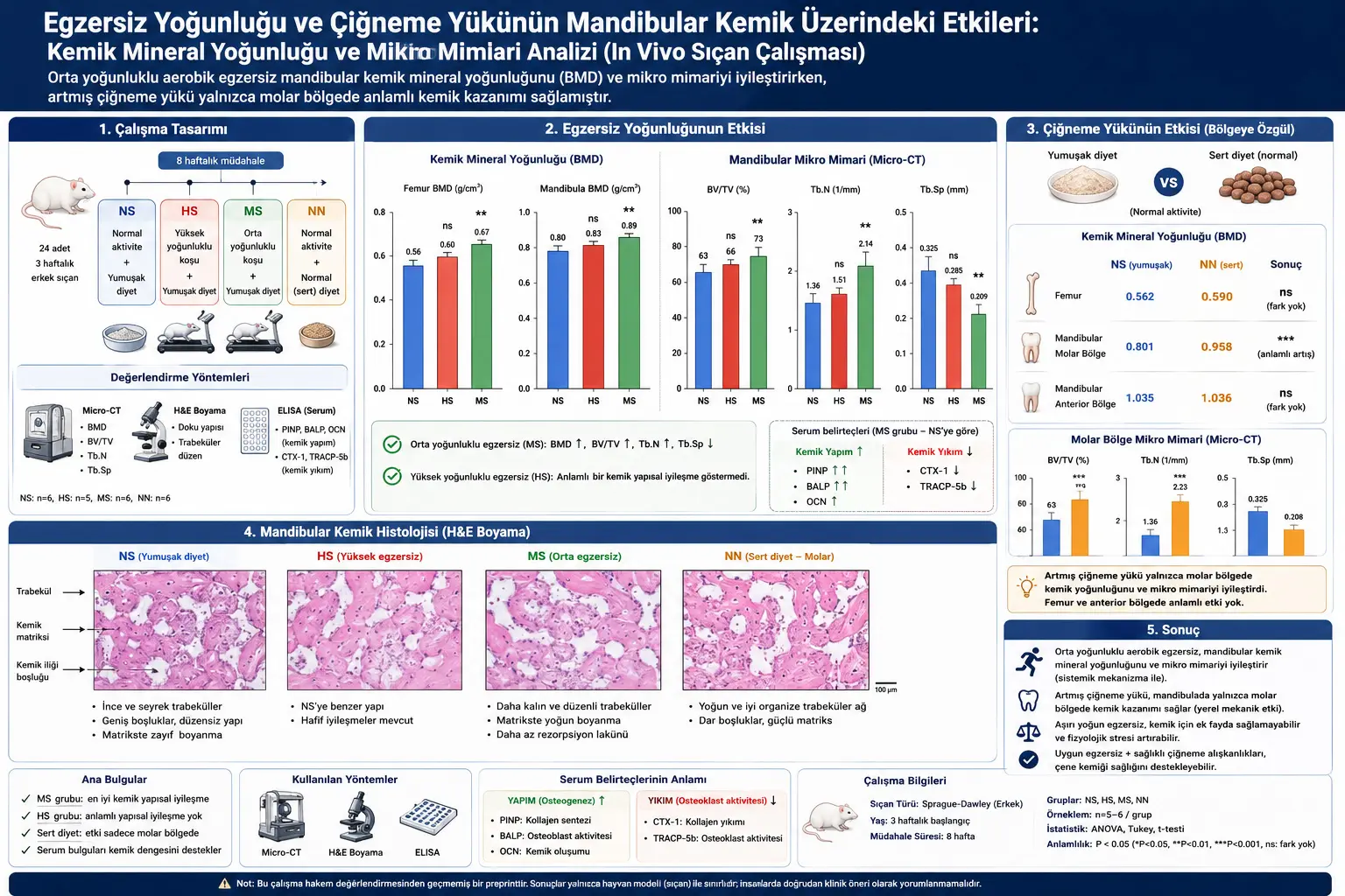
Strongest finding is positive effect of moderate-intensity exercise on both femur and mandible. Femur BMD in MS was 0,6662 ± 0,0380 g/cm³, in NS 0,5621 ± 0,0527 g/cm³. Mandibular BMD in MS was 0,8885 ± 0,0315 g/cm³, in NS 0,8013 ± 0,0360 g/cm³. Differences were statistically significant.
Mandibular microarchitecture also improved with moderate exercise. In MS BV/TV 72,8310 ± 1,7814%, Tb.N 2,1434 ± 0,4630 1/mm, Tb.Sp 0,2094 ± 0,0779 mm. In NS BV/TV 62,5270 ± 10,3051%, Tb.N 1,3598 ± 0,0618 1/mm, Tb.Sp 0,3247 ± 0,0273 mm. Thus bone volume fraction and trabecular number increased while trabecular separation decreased.
Histologically, MS group showed best appearance. Trabecular bone was more densely and regularly distributed, network structure more integrated, trabeculae more uniform in thickness, resorption lacunae rare. Bone matrix stained more densely and homogeneously, osteocytes showed more normal morphology. These findings are consistent with micro-CT results.
Чаро high-intensity exercise same effect надод?
In high-intensity group, some serum markers changed. PINP and BALP increased, TRACP-5b decreased. But OCN and CTX-1 showed no significant difference. More importantly, biochemical changes did not translate into significant gain in femur or mandibular BMD. HS femur BMD was 0,6036 ± 0,0423 g/cm³ and mandibular BMD 0,8290 ± 0,0099 g/cm³; no significant difference vs NS.
Study interprets this as biphasic effect of exercise. Moderate mechanical loading may stimulate bone formation, while overly intense or prolonged exercise may disrupt skeletal adaptation. High-intensity exercise can increase oxidative stress, stress hormones and physiological load, offsetting or reducing osteogenic benefit. Death of one rat in HS also suggests protocol may have caused substantial physiological stress.
Finding does not support simplistic inference “the more intense exercise, the better for bone.” Study says more cautiously: in examined rat model, moderate-intensity aerobic exercise appeared beneficial to mandibula, whereas high-intensity exercise under same conditions did not provide significant structural bone gain.
Masticatory load чӣ гуна effect ба mandibula showed?
Second main section evaluated masticatory load from normal-hard diet. NN got normal pellet diet, NS got same nutritional content in soft form. Thus difference intended to arise from physical hardness and chewing load, not nutrient composition.
Results were highly regional. Mandibular molar-region BMD in NN was 0,9581 ± 0,0652 g/cm³, in NS 0,8013 ± 0,0360 g/cm³, a significant difference. Femur BMD NN 0,5898 ± 0,1002 g/cm³ vs NS 0,5621 ± 0,0526 g/cm³, no significant difference. Anterior mandibular BMD NN 1,0358 ± 0,0114 g/cm³ vs NS 1,0353 ± 0,0101 g/cm³, no significant difference.
Molar-region microarchitecture also improved with hard diet. NN BV/TV 74,9742 ± 2,7114%, Tb.N 2,2326 ± 0,6379 1/mm, Tb.Sp 0,2081 ± 0,0819 mm. NS BV/TV 62,5270 ± 10,3051%, Tb.N 1,3598 ± 0,0618 1/mm, Tb.Sp 0,3247 ± 0,0273 mm. Hard diet was associated with greater bone volume, more trabeculae and less trabecular space in molar region.
Finding according to Wolff’s law чӣ маъно дорад?
Study relates masticatory-load finding to Wolff’s law. According to Wolff’s law, bone adapts to mechanical environment. In regions with increased load, bone formation may increase and bone mass strengthen; in underloaded regions, bone-loss tendency may occur.
Here normal-hard diet required greater grinding/crushing forces especially in molar region. Molar region bears longer-duration and higher-frequency load during chewing. Therefore bone adaptation was particularly evident there. Femur was not directly exposed to masticatory load and did not change. Anterior mandibular region participates in cutting/gnawing but is not main center of prolonged grinding load, so no significant change occurred.
This result shows exercise and masticatory load have different mechanisms. Exercise can create systemic effects by changing serum bone-metabolism markers and affecting distant bones like femur and mandible. Masticatory load is more local mechanical stimulus and shows stronger effect in directly loaded molar region.
Чаро anterior mandibular region unaffected буд?
Unexpected point is that hard diet did not significantly change anterior mandibular region. Small differences in BMD, BV/TV, Tb.N and Tb.Sp between NN and NS were not statistically significant.
Researchers suggest two possible reasons. First, rat incisors and molars have different functions. Incisors used for cutting/gnawing; molars for prolonged grinding/crushing. Normal-hard diet may create more load specifically during grinding phase. Therefore mechanical-stimulus threshold may be exceeded in molar region but not anterior region.
Second, rat incisors grow continuously throughout life. Because of this physiology, anterior alveolar bone is already under active remodeling. High basal activity may reduce sensitivity to additional chewing load. Study notes anterior mandibular BMD and BV/TV were higher than molar region in both NN and NS, indicating baseline bone mass differs by mandibular region.
Шаклҳо ва tables чӣ нишон медиҳанд?
BMD graphs for exercise groups визуалӣ нишон медиҳанд, ки femur ва mandibula bone mineral density дар moderate-intensity exercise group ба таври равшан зиёд шудааст, аммо дар high-intensity exercise group significant difference нест. Ин graphs main visual basis of conclusion “moderate intensity effective, high intensity ineffective” мебошанд.
Mandibular microarchitecture graphs нишон медиҳанд, ки дар MS group BV/TV ва Tb.N зиёд ва Tb.Sp кам шудааст. Аз trabecular perspective, ин маънои fuller, more connected ва less porous bone architecture-ро дорад. Micro-CT two- ва three-dimensional images numerical results-ро support мекунанд; trabecular network дар MS group more regular and compact намоён аст.
H&E staining images microscopic changes-ро нишон медиҳанд. MS group trabeculae better organized, bone matrix more homogeneous and densely stained; NS group sparser trabeculae, wider spaces and more irregular structure дорад. HS group appears closer to NS.
Serum-marker graphs нишон медиҳанд, ки moderate exercise bone-formation markers-ро increase ва bone-resorption markers-ро decrease мекунад. Ин suggests mandibular bone gain not only local imaging finding, but related to systemic shift in bone metabolism.
Masticatory-load graphs show effect of hard diet concentrated in molar region, not femur or anterior mandibular region. Molar BMD and microarchitecture parameters significantly improve with hard diet. Anterior-region graphs show “ns”, meaning nonsignificant differences. This supports region-specific masticatory-load conclusion.
Аҳамияти study аз гузашта, имрӯз ва оянда
Historically bone-health research focused more on classic weight-bearing bones such as spine, hip and femur. Jaw bone often treated as local structure in dentistry. This study connects two areas by showing mandibula may respond via different pathways to systemic exercise and local masticatory loading.
For today, finding supports idea that oral health and general body health should not be considered entirely separate. Positive effect of moderate exercise on mandibular BMD and microarchitecture suggests jaw bone may be linked to systemic bone metabolism. Аммо this is rat-model result and should not be directly converted to human clinical recommendation.
For future, study creates questions for dental implantology, periodontal bone loss, orthodontic anchorage and age-related jaw weakness. Effects of exercise may differ in aged, osteoporotic or hormonally altered animal models. In humans, relationships between chewing function, diet texture, exercise habit and mandibular bone quality need further study.
Таъсир ба daily life чӣ гуна бояд understood шавад?
Cautious practical interpretation is that bone is living, dynamic tissue responding to mechanical stimuli. Moderate regular physical activity in rat model was associated with systemic bone-metabolism changes benefiting not only weight-bearing femur but also mandibula, which does not directly bear running load.
For chewing load, study shows soft and hard diets do not have same mandibular effect. Increased chewing load from hard food strengthened bone density and trabecular structure especially around molars. Аммо this does not mean random hard-food consumption or chewing exercise should be recommended to humans. Human dentition, temporomandibular joint, periodontal disease, implants, orthodontic treatment and jaw pain vary widely.
Therefore practical meaning is not prescription but demonstration of biological principle: moderate systemic activity and local mechanical load can influence structural adaptation of bone. Clinical human studies are needed before translating this into safe/effective application.
Ҷиҳатҳои қавии study
One strength is separate testing of exercise intensities. Moderate and high intensity were not lumped together; study experimentally showed they can yield different biological outcomes.
Second strength is mandibular effects were not evaluated only by BMD. Micro-CT measured BV/TV, Tb.N and Tb.Sp; H&E evaluated histology; ELISA measured serum bone-metabolism markers. Thus conclusions rely on multiple evidence layers.
Third strength is separate comparison of masticatory load effects on femur, mandibular molar region and anterior region. This allowed understanding that local mechanical load had stronger effect where directly applied.
Маҳдудиятҳои study
Important limitations exist. First, only young male rats were used, with active bone metabolism. Generalization to older, female, osteoporotic or hormone-deficient models is uncertain.
Second, rat mandible is not identical to human mandible. Rat chewing behavior, dental structure, continuously growing incisors and mandibular anatomy differ from humans. Thus findings are interesting for dentistry but should not be presented as direct human clinical outcomes.
Third, one animal died in high-intensity group. This suggests protocol may have caused substantial physiological stress and requires caution. Larger studies could better define exercise-intensity threshold and safe ranges.
Fourth, study is preprint and not peer reviewed. Therefore results are detailed but unreviewed experimental findings, not definitive peer-reviewed evidence.
Study чӣ мегӯяд ва чӣ намегӯяд?
Study shows moderate-intensity aerobic exercise improved mandibular BMD and microarchitecture in young male rats, consistent with changes in serum bone formation/resorption markers. It also shows increased masticatory load from normal-hard diet produced local bone gain in mandibular molar region but not significant effect in femur or anterior mandible.
Study does not say same exercise protocol will strengthen human jaw bone in same way. It does not claim hard-food consumption is safe or therapeutic for everyone. It provides no direct clinical recommendation for dental implant success, periodontal disease, TMJ health or orthodontic treatment. Findings are limited to animal model and require additional human research before clinical application.
Усул ва Натиҷаҳои Таҳқиқот
Experimental design
| Group | Activity | Diet | Purpose |
|---|---|---|---|
| NS | Normal activity | Soft diet | Basic control for exercise and chewing comparisons. |
| HS | High-intensity treadmill exercise | Soft diet | Assess effect of high-intensity aerobic exercise. |
| MS | Moderate-intensity treadmill exercise | Soft diet | Assess effect of moderate-intensity aerobic exercise. |
| NN | Normal activity | Normal-hard diet | Assess regional effect of increased masticatory load. |
Analysis methods used
- Micro-CT: Used to measure BMD, BV/TV, Tb.N and Tb.Sp in femur and mandible.
- H&E staining: Mandibular trabecular structure, bone matrix, osteocyte appearance and marrow structure were examined histologically.
- ELISA: Serum PINP, BALP, OCN, CTX-1 and TRACP-5b levels measured.
- Statistics: Data presented as mean ± standard deviation; ANOVA, Tukey post hoc and independent-sample t-test used; significance threshold P < 0,05.
BMD results in exercise groups
| Group | Femur BMD (g/cm³) | Mandibula BMD (g/cm³) | Interpretation |
|---|---|---|---|
| HS | 0,6036 ± 0,0423 | 0,8290 ± 0,0099 | No significant improvement compared with NS. |
| MS | 0,6662 ± 0,0380 | 0,8885 ± 0,0315 | Significant BMD increase in femur and mandible. |
| NS | 0,5621 ± 0,0527 | 0,8013 ± 0,0360 | Soft-diet and normal-activity control. |
Mandibular microarchitecture in exercise groups
| Group | BV/TV (%) | Tb.N (1/mm) | Tb.Sp (mm) | Interpretation |
|---|---|---|---|---|
| HS | 66,4217 ± 4,8997 | 1,5097 ± 0,3034 | 0,2850 ± 0,0767 | No significant microarchitectural improvement compared with NS. |
| MS | 72,8310 ± 1,7814 | 2,1434 ± 0,4630 | 0,2094 ± 0,0779 | BV/TV and Tb.N increased, Tb.Sp decreased; trabecular structure improved. |
| NS | 62,5270 ± 10,3051 | 1,3598 ± 0,0618 | 0,3247 ± 0,0273 | Control group; weakest microarchitectural appearance. |
Serum bone-formation markers
| Group | PINP (ng/ml) | BALP (ng/ml) | OCN (ng/ml) | Interpretation |
|---|---|---|---|---|
| HS | 12,3130 ± 0,7301 | 23,0714 ± 0,7126 | 1,9298 ± 0,2808 | PINP and BALP increased but did not translate into bone-density gain. |
| MS | 13,6849 ± 1,3778 | 32,6545 ± 1,8205 | 2,2155 ± 0,1089 | Bone-formation markers clearly increased. |
| NS | 9,3260 ± 0,8586 | 26,0583 ± 1,8766 | 1,6896 ± 0,1026 | Control level. |
Serum bone-resorption markers
| Group | CTX-1 (ng/ml) | TRACP-5b (ng/ml) | Interpretation |
|---|---|---|---|
| HS | 0,8523 ± 0,0253 | 2,5355 ± 0,13612 | TRACP-5b decreased; no significant difference stated for CTX-1. |
| MS | 1,3110 ± 0,1269 | 2,1561 ± 0,1928 | Text states CTX-1 and TRACP-5b significantly decreased vs NS; however table values appear directionally inconsistent for CTX-1 and require cautious interpretation. |
| NS | 1,1951 ± 0,2390 | 3,0847 ± 0,32530 | Control level. |
Note: Source text states that CTX-1 level in MS group significantly decreased compared with NS, but table value for MS appears higher than NS. Therefore CTX-1 result should be conveyed without silently correcting this possible discrepancy between narrative and table.
Masticatory load: BMD results
| Group | Femur BMD (g/cm³) | Mandibular molar-region BMD (g/cm³) | Mandibular anterior-region BMD (g/cm³) | Interpretation |
|---|---|---|---|---|
| NS | 0,5621 ± 0,0526 | 0,8013 ± 0,0360 | 1,0353 ± 0,0101 | Soft-diet control. |
| NN | 0,5898 ± 0,1002 | 0,9581 ± 0,0652 | 1,0358 ± 0,0114 | Significant increase in molar region; no significant difference in femur or anterior region. |
Masticatory load: Molar-region microarchitecture
| Group | BV/TV (%) | Tb.N (1/mm) | Tb.Sp (mm) | Interpretation |
|---|---|---|---|---|
| NS | 62,5270 ± 10,3051 | 1,3598 ± 0,0618 | 0,3247 ± 0,0273 | Weaker molar trabecular structure with soft diet. |
| NN | 74,9742 ± 2,7114 | 2,2326 ± 0,6379 | 0,2081 ± 0,0819 | Hard diet associated with higher bone-volume fraction, more trabeculae and less trabecular spacing in molar region. |
Masticatory load: Anterior-region microarchitecture
| Group | BV/TV (%) | Tb.N (1/mm) | Tb.Sp (mm) | Interpretation |
|---|---|---|---|---|
| NS | 80,4085 ± 1,4527 | 1,1473 ± 0,0235 | 0,1728 ± 0,0169 | Soft-diet anterior-region value. |
| NN | 80,4875 ± 0,7497 | 1,1418 ± 0,0271 | 0,1627 ± 0,0111 | No significant difference despite hard diet. |
General findings table
| Intervention | Affected region | Main result | Mechanistic interpretation |
|---|---|---|---|
| Moderate-intensity aerobic exercise | Femur and mandible | BMD and mandibular microarchitecture improved. | Systemic bone metabolism may have shifted toward bone formation. |
| High-intensity aerobic exercise | Femur and mandible | No significant BMD or microarchitectural improvement. | Excess physiological stress may have limited osteogenic benefit. |
| Normal-hard diet / increased masticatory load | Mandibular molar region | BMD and trabecular structure significantly improved. | Local mechanical load may have caused regional adaptation consistent with Wolff’s law. |
| Normal-hard diet / increased masticatory load | Femur and mandibular anterior region | No significant change. | Masticatory load produced no systemic effect; anterior region may not have reached sufficient loading threshold. |
Ёддошт оид ба Манбаъ ва Усул
Ин мақола дар асоси таҳқиқоти Yiding Chang, Binyuan Gao, Jinrong Yan ва Xinrong Nan бо унвони “Effects of Aerobic Exercise Intensity and Masticatory Load on Mandibular Bone Mineral Density and Microarchitecture: An In Vivo Rat Study” таҳия шудааст. Author information presents affiliation with Shanxi Medical University School and Hospital of Stomatology.
Source text is preprint research paper on SSRN and explicitly states “This preprint research paper has not been peer reviewed”. Therefore study has not passed peer review. Findings should be read as results of experimental in vivo animal study in young male rats, not peer-reviewed clinical evidence or direct treatment recommendations.
Explanations here are based on PDF study. No claims of human clinical effect, dental implant success, jaw-bone treatment, exercise prescription, hard-food recommendation or definite health guarantee not in PDF have been added. Results are limited to animal model, micro-CT, H&E staining and ELISA measurements.
Study reports moderate-intensity exercise improved mandibular bone mineral density and microarchitecture; high-intensity exercise under same conditions did not yield significant structural bone benefit; increased masticatory load from normal-hard diet produced clear effect only in mandibular molar region. No significant chewing-load-dependent change in femur or anterior mandible was observed.
Limitations are important. Only young male rats used. Older, female, osteoporotic or hormonally different models not studied. Rat chewing pattern and mandibular anatomy differ from humans. Findings should not be directly transferred to human dentistry or general-health practice. Study is also unreviewed and needs support from larger confirmatory studies.
Шарҳ гузоред
Нишонии почтаи электронии шумо нашр намешавад. Майдонҳои ҳатмӣ бо * нишон дода шудаанд